Origin of the Mitotic Figure
spindle and centrosome
ORIGIN OF THE MITOTIC FIGURE The chromatic figure (chromosomes) is derived directly from the chromatic network of the resting-nucleus as described above. The derivation of the achromatic figure (spindle and asters) is a far more difficult question, which is still to some extent involved in doubt. By the earlier observers (1873-75) the achromatic figure was supposed to disappear entirely at the close of cell-division, and most of them (Butschli, Strasburger, Van Beneden, '75) believed it to be reformed at each succeeding division out of the nuclear substance. Later researches (1875-85) gave contradictory and apparently irreconcilable results. Fol ('79) derived the spindle from the nuclear material, the asters from the cytoplasm. Strasburger ('80) asserted that the entire achromatic figure arose from the cytoplasm. Flemming ('82) was in doubt, and regarded the question of nuclear or cytoplasmic origin as one of minor importance, yet on the whole inclined to the opinion that the achromatic figure arose inside the nucleus.' In 1887 a new face was put on the whole question through the independent discovery by Van Beneden and Boveri that the centrosome does not disappear at the close of mitosis, but remains as a distinct cell-organ lying beside the nucleus in the cytoplasm. These investigators agreed that the amphiaster is formed under the influence of the centrosome, which leads the way in cell-division by dividing into two similar halves to form the centres of division. " Thus we are justified," said Van Beneden, " in regarding the attraction-sphere with its central A. Spermatogonium in the spireme-stage; the chromatin-thread lies in the linin-network, still surrounded by the membrane ; above, the two centrosomes, the central spindle not yet formed. B. Later stage (spermatocyte) ; the nuclear membrane has disappeared, leaving the naked chromosomes; above, the amphiaster, with centrosomes and central spindle; astral rays extending towards the chromosomes. D. Following stage; splitting of the chromosomes, growth of the aster; mantle-fibres and central spindle clearly distinguished. C. The fully formed mitotic figure (metaphase) ; the chromosomes, fully divided, grouped in the equatorial plate.

corpuscle as forming a permanent organ, not only of the early blastomeres, but of all cells ; that it constitutes a cell-organ equal in rank to the nucleus itself ; that every central corpuscle is derived from a pre-existing corpuscle, every attraction-sphere from the pre-existing sphere, and that division of the sphere precedes that of the cell Boveri expressed himself in similar terms in the same year ('87, 2, p. 153), and the same general result was reached by Vejdovsky nearly at the same though it was less clearly formulated than by either Boveri or Van Beneden.
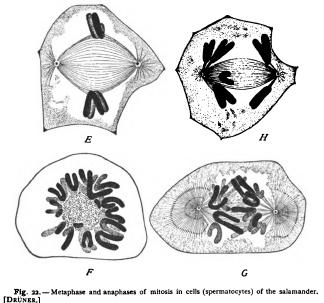
E. Metaphase. The continuous central spindle-fibres pass from pole to pole of the spindle. Outside them the thin layer of contractile mantle-fibres attached to the divided chromosomes, of which only two are shown. Centrosomes and asters. F. Transverse section through the mitotic figure showing the ring of chromosomes surrounding the central spindle, the cut fibres of the latter appearing as dots. G. Anaphase ; divergence of the daughter-chromosomes, exposing the central spindle as the interzonal fibres; contractile fibres (principal cones of Van Beneden) clearly shown. H. Later anaphase (dyaster of Flemming) ; the central spindle fully exposed to view ; mantle-fibres attached to the chromosomes. Immediately afterwards the cell divides (see Fig. 23).
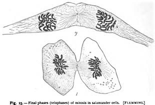
All these observers agreed, therefore, that the achromatic figure arose outside the nucleus, in the cytoplasm ; that the primary impulse to cell-division was given, not by the nucleus, but by the centrosome, and that a new cell-organ had been discovered whose special office was to preside over cell-division. " The centrosome is an independent permanent cell-organ, which, exactly like the chromatic elements, is transmitted by division to the daughter-cells. The centrosome represents the dynamic centre of the This view has been widely accepted by later investigators, and the centrosome has been shown to occur in a large number of adult tissue-cells during their resting state ; for example in pigment-cells, leucocytes, connective tissuecells, epithelial and endothelial cells, in certain gland-cells and nervecells, in the cells of many plant-tissues, and in some of the unicellular I. Epithelial cell from the lung; chromosomes at the poles of the spindle, the cell-body dividing; granules of the " mid-body " or Zwtschenk.drper at the equator of the disappearing spindle. 7. Connective-tissue cell (lung) immediately after division ; daughter-nuclei reforming, the centrosome just outside of each ; mid-body a single granule in the middle of the remains of the spindle.
plants, and animals, such as the Diatoms and Flagellates. That the centrosome gives the primary impulse to cell-division by its own division has, however, been disproved ; for there are several accurately determined cases in which the chromatin-elements divide long before the centrosome, and it is now generally agreed that the division of chromatin and centrosome are two parallel events, the causal relation between which still remains undetermined. (Cf.