
EYE, ANATOMY OF. The eye consists of the eyeball, certain muscles which move it, and the lachrymal apparatus which keeps the front of it moist. The eyeball is contained in the front of the orbit and is a sphere of about an inch (24 mm.) in diameter. From the front of this a segment of a lesser sphere pro jects slightly and forms the cornea (see fig. 1). The eyeball has three coats, external (protective), middle (vascular), and internal (sensory). There are also three refracting media, the aqueous humour, the lens and the vitreous humour or body.
The protective coat consists of the sclerotic in the posterior five-sixths and the cornea in the anterior sixth. The sclerotic or "white of the eye" (see fig. 1) is a firm fibrous coat, posteriorly pierced by the optic nerve. The cornea is continuous with the sclerotic but has a greater convexity. It consists of five layers, the outermost of which is stratified epithelium. Its transparency is due to the fact that all these layers have the same refractive index.
The middle or vascular coat of the eye consists of the choroid, the ciliary processes and the iris. The choroid (see fig. i) does not come quite as far forward as the corneo-scleral junction ; it is composed of numerous blood-vessels and pigment cells bound together by connective tissue.
The ciliary processes are some 7o triangular ridges, radially arranged, with their apices pointing backward (see fig. I), while their bases are level with the corneo-scleral junction. They are as vascular as the rest of the choroid, and contain in their in terior the ciliary muscle, which consists of radiating and circular fibres. The radiating fibres (see fig. i) pull forward the choroid when they contract. The circular fibres lie just internal to these and are few or wanting in short-sighted people.
The iris (see fig. I) is the coloured diaphragm of the eye, the centre of which is pierced to form the pupil ; it is composed of a connective tissue stroma containing blood-vessels, pigment cells and muscle fibres. The pigment in the substance of the iris is variously coloured in individuals, and is often deposited after birth, so that, in newly born European children, the colour of the eyes is often slate-blue owing to the black pigment at the back of the iris showing through. White, yellow or reddish-brown pigment is deposited later in the substance of the iris, causing the appearance, with the black pigment behind, of grey, hazel or brown eyes. In blue-eyed people very little interstitial pigment is formed, while in Albinos the posterior pigment is also absent and the blood vessels give the pink coloration. The muscle fibres of the iris are circular and radiating, but it is uncertain whether the latter are really muscular or elastic.

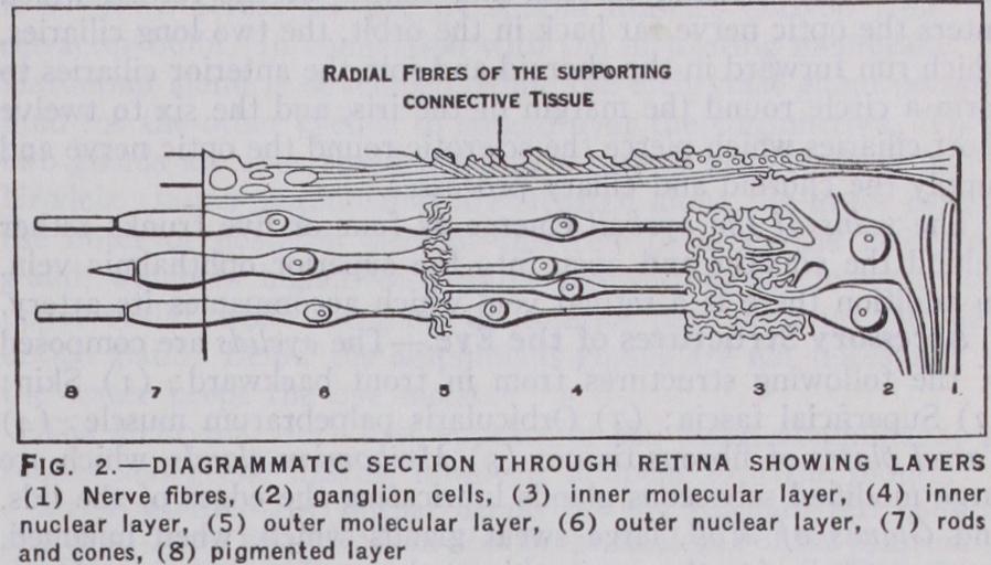
The inner or sensory layer of the wall of the eyeball is the retina; it is a delicate transparent membrane which becomes thin ner as the front of the eye is approached. A short distance be hind the ciliary processes the nervous part of it stops, and forms a scalloped border called the ora serrata. Under the microscope the posterior part of the retina is seen to consist, from front to back, of eight layers (fig. 2) as follows: (I) Layer of nerve fibres; (2) Layer of ganglion cells; (3) Inner molecular layer; (4) Inner nuclear layer ; (5) Outer molecular layer ; (6) Outer nuclear layer; (7) Layer of rods and cones; (8) Pigmented layer. Supporting the delicate nervous structures of the retina and bind ing them together is a series of radial fibres, or connective tissue rods, known as the fibres of Muller.
When the retina is looked at with the naked eye from in front two small marks are seen on it. One of these is an oval depression about 3 mm. across, which, owing to the presence of pigment, is known as the yellow spot (macula lutea) ; it is situated directly in the antero-posterior axis of the eyeball, and at its margin the nerve fibre layer is thinned and the ganglionic layer thickened. At its centre, however, both these layers are wanting, and in the layer of rods and cones only the cones are present. This central part is called the fovea centralis and is the point of acutest vision. The second mark is a little below and to the inner side of the yellow spot ; it is a circular disc with raised margins and a de pressed centre and is called the optic disc; in structure it is a complete contrast to the yellow spot, for all the layers except that of the nerve fibres are wanting, and consequently, as light cannot be appreciated here, it is known as the "blind spot." It marks the point of entry of the optic nerve, and at its centre the retinal artery appears and divides into branches. An appreciation of the condition of the optic disc is one of the chief objects of the ophthalmoscope.
The crystalline lens (see fig. i) with its ligaments separates the aqueous from the vitreous chamber of the eye; it is biconvex and the posterior surface is more curved than the anterior. Radiating from the anterior and posterior poles are three faint lines forming a y, the posterior y being erect and the anterior inverted. Running from these figures are lamellae, like the layers of an onion, each of which is made up of fibrils called the lens fibres. The whole lens is enclosed in an elastic structureless membrane, and, like the cornea, its transparency is due to the fact that all its constituents have the same refractive index.
The ligament of the lens is the thickened anterior part of the hyaloid membrane which surrounds the vitreous body; it is closely connected to the iris at the ora serrata, and then splits into two layers, of which the anterior is the thicker and blends with the anterior part of the elastic capsule of the lens, so that, when its attachment to the ora serrata is drawn forward by the ciliary muscle, the lens by its own elasticity, increases its con vexity. Between the anterior and posterior splitting of the hyaloid membrane is a circular lymph space surrounding the margin of the lens known as the canal of Petit.
The aqueous humour (see fig. i) is contained between the lens and its ligament posteriorly and the cornea anteriorly. It is prac tically a very weak solution of common salt (chloride of sodium 1.4%). The space containing it is unequally divided into a large anterior and a small posterior chamber by a perforated diaphragm —the iris.
The vitreous body or humour is a jelly which fills all the con tents of the eyeball behind the lens. It is surrounded by the hyaloid membrane, and anteriorly is concave for the reception of the lens. The composition of the vitreous is practically the same as that of the aqueous humour.
The arteries of the eyeball are derived from the ophthalmic branch of the internal carotid, and consist of the retinal which enters the optic nerve far back in the orbit, the two long ciliaries, which run forward in the choroid and join the anterior ciliaries to form a circle round the margin of the iris, and the six to twelve short ciliaries which pierce the sclerotic round the optic nerve and supply the choroid and ciliary processes.
The veins of the eyeball emerge as four or five trunks rather behind the equator and open into the superior ophthalmic vein. In addition there is a retinal vein which accompanies its artery.
The lachrymal gland is found in the upper and outer part of the front of the orbit and is about the size of an almond. Its six to twelve ducts open on to the upper reflection of the conjunctiva.
The lachrymal canals (canaliculi) (indicated in fig. 3) are superior and inferior, and open by minute orifices (puncta) on to the free margins of the two eyelids near their inner point of junction. They collect the tears, secreted by the lachrymal gland, which thus pass right across the front of the eyeball, continually moistening the conjunctiva. The two ducts are bent round a small pink tubercle (caruncula lachrymalis) (see fig. 3) at the inner angle of the eyelids, and open into the lachrymal sac (see fig. 3), which lies in a groove in the lachrymal bone. The sac is continued down into the nasal duct (see fig. 3), which is about a in. long and opens into the inferior meatus of the nose, its opening being guarded by a valve.

The orbit contains seven muscles, six of which rise close to the optic foramen. The levator palpebrae superioris is the highest, and passes forward to the superior tarsal plate and fornix of the conjunctiva. The superior and inferior recti are inserted into the upper and lower surfaces of the eyeball respectively ; they make the eye look inward as well as up or down. The external and internal recti are inserted into the sides of the eyeball and make it look outward or inward. The superior oblique runs forward to a pulley in the inner and front part of the roof of the orbit, round which it turns to be inserted into the outer and back part of the eyeball. It turns the glance downward and outward. The inferior oblique rises from the inner and front part of the floor of the orbit, and is also inserted into the outer and back part of the eyeball. It directs the glance upward and outward. Of all these muscles the superior oblique is supplied by the fourth cranial nerve, the external rectus by the sixth and the rest by the third.
The posterior part of the eyeball and the anterior parts of the muscles are enveloped in a lymph space, known as the capsule of Tenon, which assists their movements.
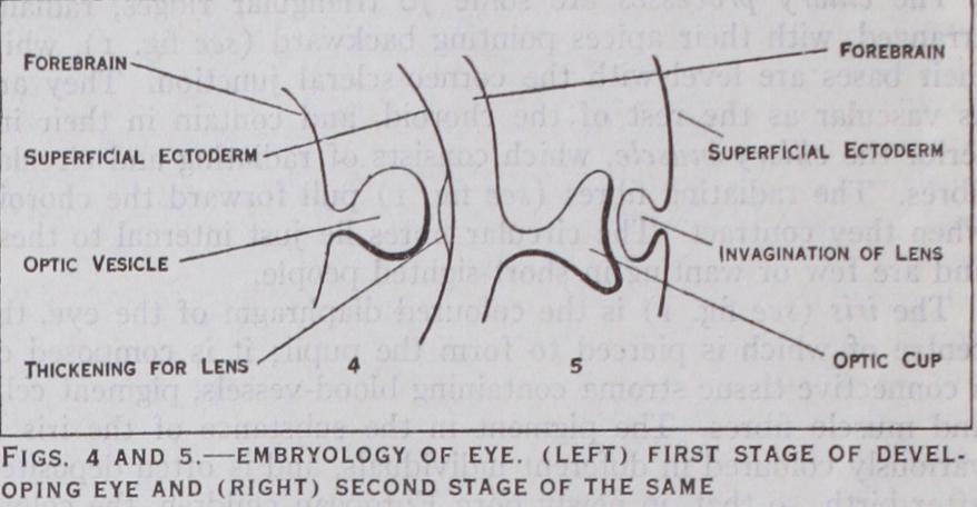
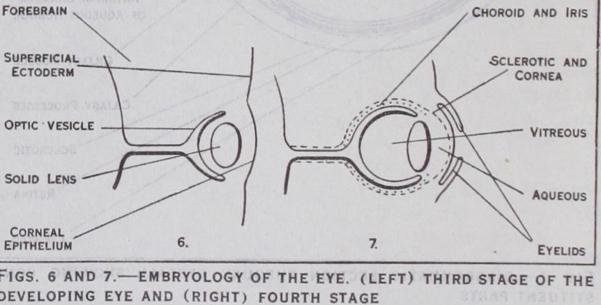
The eyelids are developed as ectodermal folds, which blend with one another about the third month and separate again before birth in man (see fig. 7). The lachrymal sac and duct are formed from solid ectodermal thickenings which later become canalized.
It will thus be seen that the optic nerve and retina are formed from the brain ectoderm ; the lens, anterior epithelium of the cornea, skin of the eyelids, conjunctive and lachrymal apparatus from the superficial ectoderm; while the sclerotic, choroid, vitre ous and aqueous humours as well as the iris and cornea are de rived from the mesoderm.
In fishes the eyeball is flattened in front, but the flat cornea is compensated by a spherical lens, which, unlike that of other vertebrates, is adapted for near vision when at rest. The iris in some bony fishes (Teleostei) is not contractile. In the Teleostei, too, there is a process of the choroid (processus falciformis) which projects into the vitreous chamber and runs forward to the lens; besides nourishing the lens it is concerned in accommoda tion. This group of fishes is also remarkable for the possession of a so-called choroid gland, which is really an arterial network between the choroid and sclerotic. The sclerotic in fishes is usually chondrified and sometimes calcified or ossified. In the retina the rods and cones are about equal in number, and the cones are very large. In the cartilaginous fishes (Elasmobranchs) there is a silvery layer (tapetum lucidurn), on the retinal surface of the choroid.
In the Amphibia the cornea is more convex than in the fish, but the lens is circular and the sclerotic often chondrified. The class shows the first rudiments of the ciliary muscle, although accom modation is brought about by shifting the lens. In the retina the rods outnumber the cones. The latter are smaller than in other animals.
In Reptilia the eye is spherical and its anterior part is often protected by bony plates in the sclerotic (Lacertilia and Che lonia). The ciliary muscle is striated, and in most reptiles accom modation is effected by relaxing the ciliary ligament as in higher vertebrates, though in the snakes (Ophidia) the lens is shifted as it is in the lower forms. Many lizards have a vascular projection of the choroid into the vitreous, foreshadowing the pecten of birds and homologous with the processus falciformis of fishes. In the retina the rods are scarce or absent.
In birds the eye is tubular, especially in nocturnal and rap torial forms ; this is due to a lengthening of the ciliary region, which is always protected by bony plates in the sclerotic. The pecten, already mentioned in lizards, is a pleated vascular pro jection from the optic disc towards the lens which in some cases it reaches. In Apteryx this structure disappears. In the retina cones outnumber rods, but are not as numerous as in reptiles.
Among the accessory structures of the eye the retractor bulbi muscle is found in amphibians, reptiles, birds and many mam mals; its nerve supply shows that it is probably a derivative of the external or posterior rectus. The nictitating membrane or third eyelid is well developed in amphibians, reptiles, birds and some few sharks; it is less marked in mammals, and in man is only represented by the little plica sesnilunaris. When functional it is drawn across the eye by special muscles derived from the retractor bulbi. In connection with the nictitating membrane the Harderian gland is developed, while the lachrymal gland secretes fluid for the other eyelids to spread over the conjunctiva. These two glands are specialized parts of a row of glands which in the Urodela (tailed amphibians) are situated along the lower eyelid; the outer or posterior part of this row becomes the lachrymal gland, which in higher vertebrates shifts from the lower to the upper eyelid, while the inner or anterior part becomes the Har derian gland. Below the amphibians glands are not necessary, as the water keeps the eye moist.
The lachrymal duct first appears in the tailed amphibians; in snakes and gecko lizards, however, it opens into the mouth.
BIBLIOGRAPHY.-For further details see any standard text-book of Bibliography.-For further details see any standard text-book of anatomy. Later literature is noticed in the catalogue of the Museum of the Royal College of Surgeons (London) . (F. G. P.)