The Cytoplasm
threads and fig
These facts show how cautious we must be in judging the appearances seen in preserved cells, and justify in some measure the hesitation with which many existing accounts of cell-structure are received. The evidence is nevertheless overwhelmingly strong, as I believe, that not only the fibrillar and alveolar formations, but also the microsomes observed in cell-structures, are in part normal structures. This evidence is derived partly from a study of the living cell, partly from the regular and characteristic arrangement of the thread-work and microsomes in certain cases. In many Protozoa, for example, a fine alveolar structure may be seen in the living protoplasm ; and Flemming as well as many later observers has clearly seen fibrillar structures in the living cells of cartilage, epithelium connective-tissue, and some other animal cells (Fig. 9). Mikosch, also, has recently described granular threads in living plant-cells.
Almost equally conclusive is the beautifully regular arrangement of the fibrillae in ciliated cells (Fig. 13, Engelmann), in muscle-fibres and nerve fibres, and especially in the mitotic figure of dividing-cells (Figs. t6, 24), where they are likewise more or less clearly visible in life. A very convincing case is afforded by the pancreas-cells of Na-turns, which Mathews has carefully studied in my laboratory. Here the thread-work consists of long, conspicuous, definite fibrillx, some of which may under certain conditions be wound up more or less clearly in a spiral mass to form the so-called i'Vebenkern. In all these cases it is impossible to regard the thread-work as an accidental coagulation-product. On the whole, therefore, it is probable that careful treatment by reagents gives at least an approximately true picture of the normal thread-work, though we must always allow for the possible occurrence of artificial products.

The centre of the cell is occupied by a large vacuole, filled with a watery liquid. The cytoplasm forms a very regular and distinct reticulum with scattered microsomes which become very large in the peripheral zone. The larger pale bodies, lying in the ground-substance, are excretory granules (i.e. metaplasm). The nucleus, at the right, is surrounded by a thick chromatic membrane, is traversed by a very distinct linin-network, contains numerous scattered chromatingranules, and a single large nucleolus within which is a vacuole. Above are two isolated nuclei showing nucleoli and chromatin-granules suspended on the linin-threads.
One of the most beautiful
forms of cyto-reticulum with which I am acquainted has been described by Bolsius and Graf in the nephridial cells of leeches as shown in Fig. 14 (from a preparation byDr. Arnold Graf). The reticulum is here of great distinctness and regularity, and scattered microsomes are found along its threads. It appears with equal clearness, though in a somewhat different form, The nucleus contains a single intensely chromatic nucleolus, and a paler linin-network with rounded chromatin-granules. The cytoplasmic fibrillae are faintly shown passing out into the nerve-process below. (they are figured as far more distinct by Flemming.) The dark cytoplasmic masses are the deeply staining " chromophilic granules" (Nissl) of unknown function. (The centrosome, which lies near the centre of the cell, is shown in Fig. 7, C) At the left, two connective tissue-cells.
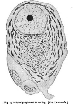
in many eggs, where the meshes are rounded and often contain foodmatters or deutoplasm in the inter-spaces (Figs. 42, 43). In cartilagecells and connective tissue-cells, where the threads can be plainly seen in life, the network is loose and open, and appears to consist of more or less completely separate threads (Fig. 9). In the cells of columnar epithelium, the threads in the peripheral part of the cell often assume a more or less parallel course, passing outwards from the central region, and giving the outer zone of the cell a striated appear-. ance. This is very conspicuously shown in ciliated epithelium, the fibrillae corresponding in number with the cilia as if continuous with their bases (Fig. In nerve-fibres the threads form closely set parallel fibrill which may be traced into the body of the nerve-cell ; here, according to most authors, they break up into a network in which are suspended numerous deeply staining masses, the " chromophilic granules" of Nissl (Fig. 15). In the contractile tissues the threads are in most cases very conspicuous and have a parallel course. This is clearly shown in smooth muscle-fibres and also, as'Ballowitz has shown, in the tails of spermatozoa. This arrangement is most striking in striped muscle-fibres where the fibrillae are extremely well marked. According to Retzius, Carnoy, Van Gehuchten, and others, the meshes have here a rectangular form, the principal fibrillw having a longitudinal course and being connected at regular intervals by transverse threads ; but the structure of the muscle-fibre is probably far more complicated than this account would lead one to suppose, and opinion is still divided as to whether the contractile substance is represented by the reticulum proper or by the ground-substance.