The Spermatozoon
middle-piece and body
THE SPERMATOZOON Although spermatozoa were among the first of animal cells observed by the microscope, their real nature was not determined for more than two hundred years after their discovery. Our modern knowledge of the subject may be dated from the year 1841, when Kolliker proved that they were not parasitic animalcules, as the early observers supposed, but the products of cells pre-existing in the parent body. Kolliker, however, did not identify them as cells, but believed them to be of purely nuclear origin. We owe to SchweiggerSeidel and La Valette St. George the proof, simultaneously brought forward by these authors in that the spermatozoon is a complete cell, consisting of nucleus and cytoplasm, and hence of the same morphological nature as the ovum. It is of extraordinary minuteness, being in many cases less than 1/100000 the bulk of the ovum.' Its precise study is therefore difficult, and it is not surprising that our knowledge of its structure and origin is still far from complete.

Flagellate Spermatozoa
In its more usual form the animal spermatozoon resembles a minute, elongated tadpole, which swims very actively about by the vibrations of a long, slender tail morphologically comparable with a single cilium or flagellum. Such a spermatozoon consists typically of four parts, as shown in Fig. 47 : I. The nucleus, which forms the main portion of the " head," and consists of a very dense and usually homogeneous mass of chromatin staining with great intensity with the so-called " nuclear dyes " (e.g. hwmatoxylin or the basic anilines such as methyl-green). It is surrounded by a very thin cytoplasmic envelope.
2. A minute apex, or apical body, as a rule of cytoplasmic origin, though apparently derived in some cases from the nucleus. This lies at the front end of the head, and in some cases terminates in a sharp spur by means of which the spermatozoon bores its way into the ovum.
3. The or connecting piece, a larger cytoplasmic body lying behind the head and giving attachment to the tail. This body shows the same staining-reaction as the tip, having an especial affinity for "plasma-stains" (acid fuchsin, etc.).
4. The tail, orflagellum, in part, at least, a cytoplasmic product developed from or in connection with the " archoplasm " (attraction-sphere or " Nebenkern ") of the mother-cell. It consists of a fibrillated axial filament surrounded by an envelope which sometimes shows a fibrillar structure, sometimes winds spirally about the axial filament, and is in certain cases differIn the sea-urchin, Toxopneustes, I estimate its bulk as being between 1/400000 and 1/30000the volume of the ovum. The inequality is in many cases very much greater.
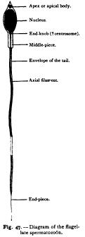
entiated into a fin-like undulating membrane. The axial filament may be traced through the middle-piece up to the head, at the base of which it terminates in a minute body, single or double, known as the end-knob, and not improbably representing the centrosome.
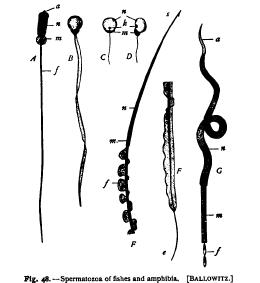
There is still some doubt regarding the nature and functions of these various parts. The nucleus is proved both by its origin and by its history during fertilization to be exactly equivalent to the nucleus of the mature egg. The middle-piece and the tail represent A. Sturgeon. B. Pike. C. D. lensicus. E. Tilton (anterior part). F. Triton (posterior part of flagellum). G. Raja (anterior part). a. apical body; e. end-piece; j. flagellum ; A. endknob (? centrosome) ; m. middle-piece ; a. nucleus; s. apical spur.
the principal mass of the cytoplasm of the sperm-cell, and the middle-piece is probably to be regarded as merely the thickened basal portion of the flagellum.
The principal uncertainty relates to the position of the centrosome. It is certain that in most cases the centrosome or attractionsphere lies in the middle-piece; for from it the centrosome arises during the fertilization of the egg, in every accurately known case. In a few cases, moreover, the middle-piece has been traced back to the attraction-sphere of the mother-cell, from which the spermatozoon is formed in the testis. On the other hand, a few observers have maintained, apparently on good evidence, that the centrosome lies, not in the middle-piece, but at the apex (p. 123).