The Spermatozoon
attached and cytoplasm
3.
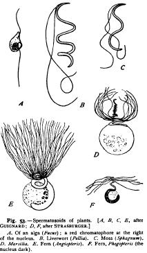
Paternal Germ-cells of Plants In the flowering plants the male germ-cell is represented by a "generative nucleus," together with two centrosomes and a small amount of cytoplasm, lying at the tip of the pollen-tube ( Fig. So, A). On the other hand, in a large number of the lower plants (Pteridophytcs, Muscinew, and many others), the male germ-cell is a minute actively swimming cell, known as the spermatozoid, which is closely analogous to the spermatozoon. The spermatozoids are in general less highly differentiated than spermatozoa, and often show a distinct resemblance to the asexual swarmers or zoospores so common in the lower plants (Figs. 52, 53). They differ in two respects from animal spermatozoa; first in possessing not one but two or several flagella ; second, in the fact that these are attached as a rule not to the end of the cell, but on the side. In the lower forms plastids are present in the form of chromatophores, one of which may be differentiated into a red " eye-spot,' as in Volvox and Fucus(Figs. 41, 53, A), and they may even contain contractile vacuoles (Volvox); but both these structures are wanting in the higher forms. These consist only of a nucleus with a very small amount of cytoplasm, and have typically a spiral form. In Chara, where their structure and development have recently been carefully studied by Belajeff, the spermatozoids have an elongated spiral form with two long flagella attached near the pointed end which is directed forwards in swimming (Fig. 52). The main body of the spermatozoid is occupied by a dense, apparently homogeneous nucleus surrounded by a very delicate layer of cytoplasm. Behind the nucleus, lies a granular mass of cytoplasm, forming one end of the cell, while in front is a slender cytoplasmic tip to which the flagella are attached. Nearly similar spermatozoids occur in the liverworts and mosses. In the ferns and other pteridophytes a somewhat different type occurs (Fig. 53). Here the spermatozoid is twisted into a conical spiral and bears numerous cilia attached along the upper turns of the spire. The nucleus occupies the lower turns, and attached to them is a large spheroidal cytoplasmic mass, which may, however, be cast off when the spermatozoid is set free or at the time it enters the archegonium. This, according to Strasburger, probably corresponds to the basal cytoplasmic mass of Chara. The upper portion of the spire to which the cilia are attached is composed of cytoplasm alone, as in Chara.
The homologies, or rather analogies, between the respective parts of the spermatozoid and spermatozoon are not yet very definitely established, since the history of the spermatozoid in fertilization has not yet been accurately followed. Strasburger ('92) believes that the anterior cytoplasmic region, to which the cilia are attached, consists of " kinoplasm " (archoplasm), and hence corresponds with the middle-piece of the spermatozoon. If this view be correct, there is, on the whole, a rather close correspondence between spermatozoid and spermatozoon, the flagella being attached in both cases to that end of the cell which contains the centrosome or kinetic centre, the nucleus lying in the middle, while the opposite end consists of cytoplasm (i.e. the apex of the spermatozoon, the cytoplasmic vesicle of pteridophytes, the basal cytoplasm of Chara, etc.). The attachment of the flagella in both cases to the archoplasmic region is a significant fact, for Strasburger believes that they arise from the " kinoplasm " (archoplasm), and it is probable that the spermatozoon tail has a similar origin (p. 126).