Modifications of Mitosis
spindle and fig
MODIFICATIONS OF MITOSIS The evidence steadily accumulates that the essential phenomena of mitosis are of the same general type in all forms of cells, both in plants and in animals. Everywhere, with a single important exception (maturation), the chromatin-thread splits lengthwise throughout its whole extent, and everywhere an achromatic spindle is formed that is in some manner an agent in the transportal of the chromatinhalves to the respective daughter-cells. The exception to this general law, which occurs during the preparation of the germ-cells for their development and constitutes one of the most significant of all cytological phenomena, is considered in Chapter V. We have here only to glance at a number of modifications that affect, not the essential character, but only the details of the typical process.
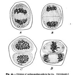
t. Varieties of the Mitotic Figure All of the mitotic phenomena, and especially those involved in the history of the achromatic figure, are in general most clearly displayed in embryonic cells, and especially in the egg-cell' (Fig. 24). In the adult tissue-cells the asters are relatively small, the spindle relatively large and conspicuous. The same is true of plant-cells in general where the very existence of the asters was at first overlooked. Plant-mitoses are characterized by the prominence of the cell-plate (Fig. 25), which is rudimentary or often wanting in animals, a fact correlated no doubt with the greater development of the cell-membrane in plants. With this again is correlated the fact that division of the cell-body in animal-cells generally takes place by constriction in the equatorial plane of the spindle ; while in plantcells the cell is usually cut in two by a cell-wall developed in the substance of the protoplasm and derived in large part from the cellplate.
The centrosome and centrosphere appear to present great variations that have not yet been thoroughly cleared up and will be more critically discussed They are known to undergo extensive changes in the cycle of cell-division and to vary greatly in different forms (Fig. 108). In some cases the aster contains at its centre nothing more than a minute deeply staining granule, which doubtless represents the centrosome alone. In other cases the granule is surrounded by a larger body, which in turn lies within the centrosphere or attraction-sphere. In still other cases the centre of the aster is
occupied by a large reticular mass, within which no smaller body can be distinguished (e.g. in pigment-cells); this mass is sometimes called the centrosome, sometimes the centrosphere. Sometimes, again, the A. Closing prophase, the equatorial plate forming. /?. Metaphase ; equatorial plate established and the chromosomes split ; b, the equatorial plate, viewed en face, showing the four chromosomes. C. Earl? anaphase; divergence of the (polar body at one side). D. Later anaphase; p.b., second polar body.

(For preceding stages sec Fig. 65; for later stages, Fig. ro4.) spindle-fibres are not focussed at a single point, and the spindle appears truncated at the ends, its fibres terminating in a transverse row of granules (maturation-spindles of Ascaris, and some plant-cells). It is not entirely certain, however, that such spindles observed in preparations represent the normal structure during life.' I Hacker asserts in a recent paper ('94) that the truncated polar spindles are normal, and that a centrosome lies at each of the four i.e. two at either pole.
The variations of the chromatic figure must for the most part be considered in the more special parts of this work. There seems to be doubt that a single continuous spireme-thread may be formed (cf. p. 184), but it is equally certain that the thread may appear from the beginning in a number of distinct segments ; i.e. as a segmented spireme. The chromosomes, when fully formed, vary greatly in appearance. In many of the tissues of adult plants and animals A. Anaphase of the first division, showing the twelve daughter-chromosomes on each side, the interzonal fibres stretching between them, and the centrosomes, already double, at the spindlepoles. B. Later stage, showing the cell-plate at the equator of the spindle and the daughterspiremes (dispireme stage of Flemming). C. Division completed ; double centrosomes in the resting cell. D. Ensuing division in progress ; the upper cell at the close of the prophases, the chromosomes and centrosomes still undivided ; lower cell in the late anaphase, cell-plate not yet formed.