General Sketch of the Ovum
polar and centrosome
The earliest investigators of fertilization, such as Butschli and Fol, had no knowledge of the centrosome, and hence no clear idea as to the origin of the asters, but Fol stated in 1873 that the asters represented " centres of attraction " lying outside and independent of the nucleus. Oscar Hertwig showed, in 1875, that in the sea-urchin egg the amphiaster arises by the division of a single aster that first appears near the sperm-nucleus and accompanies it in its progress toward the egg-nucleus. A similar observation was soon afterwards made by Fol ('79) in the eggs of Asterias and Sagitta, and in the latter case he determined the fact that the astral rays do not centre in the nucleus, as Hertwig described, but at a point in advance of it, — a fact afterwards confirmed by Hertwig himself and by Boveri ('88, 1). Hertwig and Fol afterwards found that in cases of polyspermy, when several spermatozoa enter the egg, each sperm-nucleus is accompanied by an aster, and Hertwig proved that each of these might give rise to an amphiaster (Fig. 75).
It was Boveri ('87) who first accurately traced the complete history of the centrosome and clearly formulated the facts, proving that in Ascaris a single centrosome is brought in by the spermatozoon and that it divides to form two centres about which are developed the two A. Soon after the entrance of the spermatozoon, showing the minute sperm-nucleus at ., the germinal vesicle disappearing, and the first polar mitotic figure forming. The empty spaces represent deutoplasm-spheres (slightly swollen by the reagents), the firm circles oil-drops. B. Spermnucleus (d) advancing, a minute amphiaster in front of it ; first polar mitotic figure established ; polar concentration of the protoplasm. C. Later stage ; second polar body forming. D. The polar bodies formed ; conjugation of the germ-nuclei ; the egg-centrosomes and asters have disappeared, leaving only the sperm-amphiaster (cf. Fig. 64).

asters of the cleavage-figure. He was thus led to the following conclusion, which I believe still accurately expresses the truth : "The ripe egg possesses all of the organs and qualities necessary for division excepting the centrosome, by which division is initiated. The spermatozoon, on the other hand, is provided with a centrosome, but lacks the substance in which this organ of division may exert its activity. Through the union of the two cells in fertilization all of the essential organs necessary for division are brought together; the egg now contains a centrosome which by its own division leads the way in the embryonic development.' Boveri did not actually follow the disappearance of the egg-centrosome, but nearly at the same time this process was carefully described by Vejdovsky in the case of a fresh-water annelid Rhynchelmis. Here, again, very strong evidence was brought for
A. Sperm-nucleus soon after entrance, the sperm-aster dividing. B. The germ-nuclei approaching; cr, the enlarged sperm-nucleus with a large aster at each pole; y, the egg-nucleus reformed after formation of the second polar body, shown at the right. C. The apposed reticular germ-nuclei, now of equal size; the spindle is immediately afterwards developed between the two enormous sperm-asters; polar body at the left.
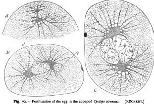
ward to show that the cleavage-amphiaster arises by the division of a single sperm-aster. Very numerous observations to the same effect have been made by later observers. Bohm could find in Petromyzon ('88) and the trout (90 no radiations near the egg-nucleus after the formation of the polar-bodies, while a beautiful sperm-aster is developed near the sperm-nucleus and divides to form the amphiaster. Platner ('86) had already made similar observations in the snail Anion, and the same result was soon afterwards reached by Brauer ('92) in the case of Branchipus, and by Julin ('93) in Styleopsis. Fick's careful study of the fertilization of the axolotl ('93) proved in a very convincing manner not only that the amphiaster is a product of the sperm-aster, but also that the latter is developed about the middle-piece as a centre. The same result was indicated by Foot's observations on the earthworm (94), and it was soon afterwards conclusively demonstrated in echinoderms through the independent and nearly simultaneous researches of myself on the egg of Toxopneustes, of Mathews on Arbacia, and of Boveri on Echinus. Nearly at the same time a careful study was made by Mead ('95) of the annelid Chaeopterus, and of the starfish Asterias by Mathews, both observers independently showing that the polar spindle contains distinct centrosomes, which, however, degenerate after the formation of the polar bodies, their place being taken by the sperm-centrosome, which divides to form an amphiaster before union of the nuclei, as in Rhynchelmis. Exactly the same result has since been reached by Hill ('95) in Sphcaerechinus and the tunicate Phallusia, and by Kostanecki and Wierzejski ('96) in Physa (Fig. 64) ; and in all of these the centrosome is likewise shown to arise from the middle-piece. The origin of the centrosome from the spermatozoon alone has also been shown by Ruckert ('95, 2) in Cyclops (Fig. 72), and is indicated by Sobotta's work ('95) on the fertilization of the mouse (Fig. 67).